Media Release: Discovery makes the invisible visible
La Trobe University researchers have led a four-year collaboration to make “the invisible visible” by using custom-designed nanomaterials to enhance the sensitivity of phase contrast microscopy, an imaging technique commonly used by scientists to study biological specimens.
The discovery, detailed in Nature photonics, will benefit a broad range of researchers and has the potential to advance research into the understanding and detection of disease.
Project leader and La Trobe Institute for Molecular Science (LIMS) physicist, Professor Brian Abbey, said the discovery allows scientists to detect minute changes in the composition or structure of transparent or nano-thin objects, enabling their key features and structures to be visible when put under a microscope.
“Features that were previously impossible to detect using conventional techniques can now be imaged using our microscopy method, developed by lead author and La Trobe Senior Research Fellow Dr Eugeniu Balaur,” Professor Abbey said.
“It has allowed our teams to delve deeper into the identification of early destructive processes in neurodegenerative diseases, such as multiple sclerosis,” Professor Abbey said.
“This technique drastically increases the contrast of sample allowing to visualize characteristic without need for staining,” says Professor Eric Hanssen, Manager of the Ian Holmes Imaging Centre at Bio21 Institute, University of Melbourne, who collaborated on this research.
Phase contrast microscopy, an optical technique that can be utilised to produce high-contrast images of transparent specimens, was first invented in 1934 by Nobel-prize winning physicist Frits Zernike.
“This technique allows scientists to examine cells in their natural state without previously being stained or labelled. As a result, their structure and function – and perhaps even their dynamics – can be better understood,” Professor Abbey said.
“We now have the tools to manipulate matter at the nanoscale. Our custom designed nanomaterials have enabled us to achieve a huge leap forward in terms of image quality and contrast, building on the groundbreaking phase work of Zernike in the 1930s.”
La Trobe neuroscientist and study co-author Dr Jacqueline Orian said the research team is working to translate the study to ensure their new microscopy technique can be utilised by scientists worldwide.
“A major application of the technique would be in the evaluation of candidate drugs for promotion of repair to nerves and their protective casings known as myelin,” Dr Orian said.
“The technique the team developed enables us to perform label-free imaging of cellular structures with exceptional contrast and in their natural state. Coupling nanotechnology with phase contrast microscopy lays the foundation for entirely new fields of research and represents a significant leap forward for the life sciences," Dr Nicholas Anthony, study co-author from the Istituto Italiano di Tecnologia (IIT) said.
The project was conducted in collaboration with researchers from the Peter McCallum Cancer Centre, the University of Melbourne, the Australian National University and IIT. Device development and fabrication was carried out at the Melbourne Centre for Nanofabrication (MCN) in the Victorian Node of the Australian National Fabrication Facility (ANFF).
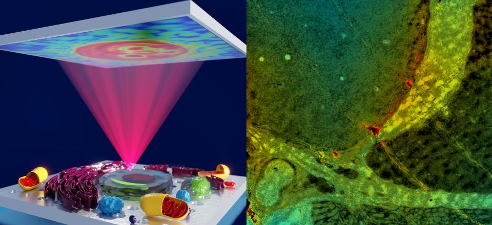
IMAGE: Concept art showing plasmon-enhanced phase microscopy. This new method will enable ultra-sensitive imaging of highly optically transparent objects in fields ranging from materials science to medical imaging
Media Contact, La Trobe University: Dragana Mrkaja – d.mrkaja [at] latrobe.edu.au – 0447 508 171
Media Contact, Bio21 Institute, University of Melbourne: Florienne Loder - florienne.loder [at] unimelb.edu.au - 0404 230 006