CryoEM insights into the nanomachine that powers the cell’s garbage disposal system

Capitalising on the revolution brought about by structural cryo-electron microscopy (cryo-EM), a Bio21 team led by Dr Mike Griffin and Prof Leann Tilley has generated molecular movies of a sophisticated nano-machine, called the proteasome.
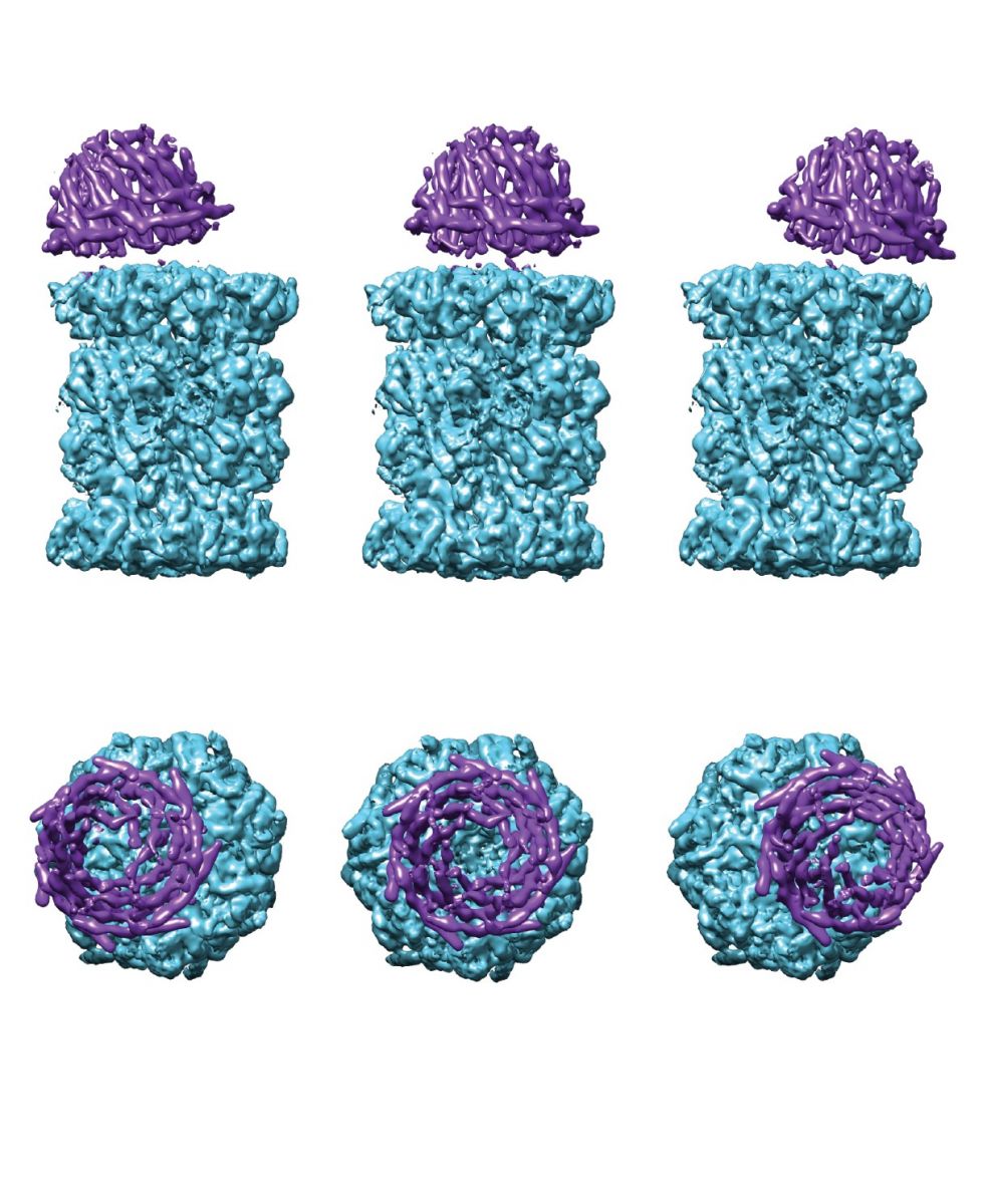
The proteasome is the cell’s waste disposal system. They showed that the barrel-shaped proteasome has a cap, called PA28, that “dances” back and forth, opening and closing a gap at the interface.
“We refer to this as the dancing proteasome motion” says Prof Tilley “and we think it opens an escape route to let shredded protein pieces out of the proteasome ready for recycling”.
S2 Single Cap Multibody from Faculty of MDHS on Vimeo.
 Figure: Dancing proteasomes. Reconstructed multibody-refined cryoEM densities showing the “dancing” of the PA28 cap (purple) on the proteasome core (aqua).
Dr Griffin explains that “Some cells such as cancerous blood cells make proteins at a gangbusters’ rate, creating so much waste they are particularly reliant on their proteasome. As a consequence, proteasome inhibitors can be used clinically as anti-cancer agents. Similarly, malaria parasites multiply rapidly inside human red blood cells and in doing so, generate a lot of waste protein. Proteasome inhibitors are under development in Prof Tilley’s lab as potential antimalarial drugs.”
To understand how living matter works, it helps to have detailed structural views of the molecular machinery. It is now possible to obtain that information, due to a revolution in structural biology, called cryo electron microscopy (cryoEM) – a revolution that was recognised by the 2017 Nobel Prize in Chemistry.
As Dr Griffin explains, “for the first time, we are able to directly image the internal mechanics of molecular machines – to see life, itself, in action.”
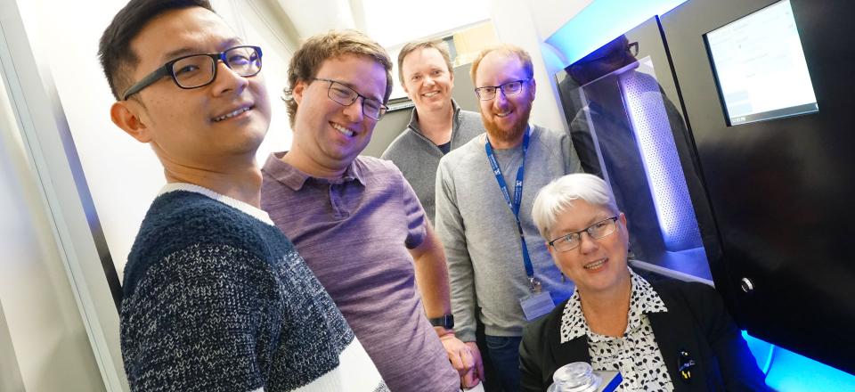
The Bio21 Institute recently established a cryoEM capability in its Melbourne Microscopy Facility and the team wanted to use it to collect the first-ever views of a proteasome working with a PA28 cap. But first they needed to purify the proteasome. Malaria parasites are only one twentieth the diameter of a human hair, so Dr Stanley Xie and Dr Tuo Yang used parasites generated during many months of culture to get enough pure protein for cryoEM studies. A heroic effort, but it was worth it.
A/Prof Eric Hanssen and Dr Andrew Leis of Bio21’s Advanced Microscopy Facility collected the cryoEM data. Putting together many thousands of electron microscopy images, a picture PA28 emerged. It looked like a conical lid sitting at the end of the proteasome barrel. The team called on the structural biology expertise of Mr Riley Metcalfe to solve the structure. At the top of the cap, flexible streamer-like loops form dynamic swirls.
As Riley explains, “we could also see loops at the bottom of PA28 and we could see how they engage with the surface of the barrel. This clasp mechanism opens up a pore in the top of the barrel, creating a channel through to the shredding enzymes in the barrel core.”
Craig Morton, Bio21 Institute, and Michael Kuiper, CSIRO Data61, used computer-based simulations to show that the dancing motion could let small peptide products escape through the interface between the cap and the barrel – providing short-cut access to and from the shredder.
Schematic illustrating how no-longer-needed proteins are fed into the proteasome barrel. Proteins are degraded and then released through the PA28 activator. The dynamic nature of the interface between PA28 and the proteasome may enable short-cut egress of peptide products.
The team’s work has been published in Nature Microbiology. Their cryoEM-based molecular movies provide new insights into the mechanism of action of this important waste disposal system. Importantly, the new high-resolution structure of the malaria parasite proteasome is also helping the design of inhibitors that specifically target the plasmodium proteasome. New antimalarial drugs are desperately needed to prevent the more than 400,000 deaths caused each year by the malaria parasite.
The team’s cryoEM structure will facilitate the rational design of new inhibitors.
Bio21 Institute is available for comment.
Media office | 03 8344 4123 | news [at] media.unimelb.edu.au ()

